
CyTOF VS Flow cytometry which method is better?

CyTOF(Mass Cytometry) VS FACS
which method is better?
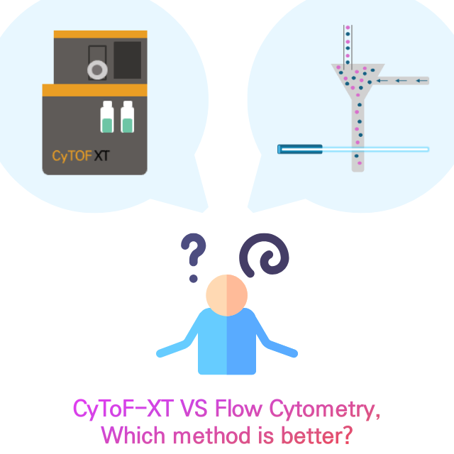
CyTOF-XT and FACS (Fluorescence-Activated Cell Sorting) are the technology for analyzing and sorting individual cells based on their properties. While they share some similarities, they also have key differences. In this article, we will look though the chracteristics of each tehcnology deeply.
What is CyToF-XT?
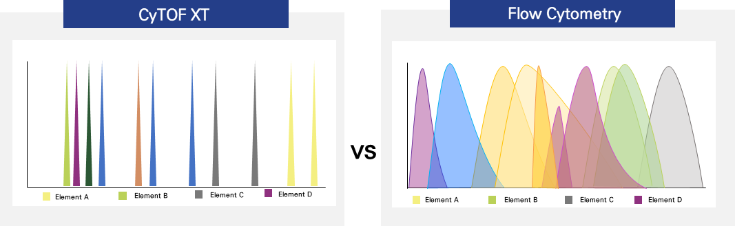
CyToF-XT, which is also known as mass cytometry is s a novel technology similar to flow cytometry in which antibodies are tagged with heavy metal molecules rather than fluorophores and then detected with time-of-flight mass spectrometry. Therefore, it reduces the spilll-over and prevent the human error by minimizing signal variability.
On the other hand, flow cytometry is a technology that provides rapid multi-parametric analysis of single cells in solution. It use lasers as light sources to produce both scattered and fluorescent light signals that are interpreted by detectors such as photodiodes or photomultiplier tubes. These light signals are converted into electronic signals that could be analyzed by a computer.
The biggest different point bewteen two method is that the material is used for the tagging. CyToF XT uses heavy metal molecules. However, flow cytometry uses a flourscent light signal. We can check the result through below image.
The usability of CyToF XT & FACS

Flow cytometry obtains a wealth of techniques and applications that are suitable for multiple fields of study. CyToF XT could be also used for below areas.
Immunophenotyping
Immunophenotyping stands as the primary application within flow cytometry, capitalizing on its distinctive capability to concurrently assess mixed cell populations across various parameters. In its basic configuration, an immunophenotyping study involves cells stained with fluorochrome-conjugated antibodies, specifically aimed at antigens present on the cell surface. These antigens are often designated with "cluster of differentiation" (CD) numbers by the Human Leukocyte Differentiation Workshops, establishing a standardized nomenclature for monoclonal antibodies targeting specific cellular antigens.
Molecular Biology
Analysis of Fluorescent Proteins Fluorescent proteins (e.g., GFP, mCherry, YFP, mRuby) serve as markers for protein expression. Typically, cells are transfected with a plasmid containing a promoter sequence and encoding a gene of interest along with a fluorescent protein. The expression of the fluorescent protein indicates the expression of the gene of interest. Recently, split bi- or tri-partite fluorescence complementation linked to other proteins has enabled the detection of RNA-protein and protein-protein interactions. These methods have transformed cell detection and isolation, where fluorescence is detected only in response to surrogates (Han et al., 2014). This technology finds applications in various areas, such as in vivo tracking of transplanted cells, detecting bacterial or viral infections, and gene knockout in cells to further understand gene function.
Cell Cycle Analysis
Cell cycle analysis assays involve staining DNA with a saturating amount of DNA-binding dye. Typically, cells are fixed with a 70% ethanol solution to permeabilize them before staining with dyes like PI, 7AAD, or DAPI. However, certain dyes can penetrate living cells and stain DNA without causing harm, such as Hoescht 33342. In this analysis, samples are acquired at a low flow rate with linear amplification and then analyzed using ploidy modeling software to determine the phases of the cell cycle.
Signal Transduction
Flow Cytometry employs antibodies generated against resting and phosphorylated signaling molecules. By using these reagents and specialized buffers in staining panels, the study of signaling pathways in mixed cell populations becomes feasible.
RNA Flow Cytometry
RNA flow cytometry combines flow cytometry with fluorescent in situ hybridization (FISH) to detect RNA expression alongside protein expression. This technique requires optimization of staining panels since not all fluorochrome-conjugated antibodies can withstand treatment at 40°C for multiple 1-hour incubations. It proves useful when antibodies are unavailable for a target, and RNA expression can be used as an alternative marker.
Cell sorting
Cell sorting employs a flow cytometer equipped with sorting capabilities to segregate and purify cells or particles for subsequent analysis. Essentially, any cell or particle that can be rendered fluorescent can be sorted using a cell sorter. Cells can be sorted into various containers such as 96 or 384 well plates, tubes, and slides. Common sample types include transfected cells expressing fluorescent proteins, stem cells, tumor-infiltrating lymphocytes, tumor cells, and white blood cell populations. An important factor to consider during cell sorting is the scalability of the antibody amount required for staining large quantities of cells.
Which method is better?
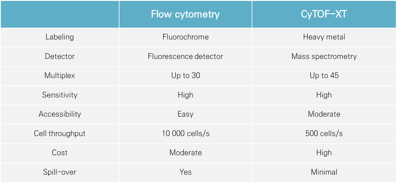
Generally, Flow cytometry has accumulated fluent data and analysis history could be used for future study. Therefore, a lot of pharmaceutical companies prefer to use flow cytometry to minimize the risk, especially for the clinical trials. However, there are some ares that could not be covered by flow cytometry, such as receptor occupancy.
The measurement of receptor occupancy, which represents the ratio of the drug bound to the total receptor on individual cells, serves as a biomarker for evaluating the response to therapeutic monoclonal antibodies. Traditionally assessed through flow cytometry, receptor occupancy faces limitations due to spectral overlap, constraining the number of markers that can be concurrently measured. Consequently, receptor occupancy assays are primarily limited to the analysis of major cell types, despite the therapeutic potential of rare cell populations. To address this, we have devised a receptor occupancy assay tailored for mass cytometry. Moreover, CyToF-XT has become more and more used based on the belief that it can cover the weakeness of flow cytometry.
Huscience is a central lab specialized in immunology and first company in south korea adopted CyToF-XT on analysis field. If you need CyTOF-XT analysis service, start the conversation with us.
Reference
[1]Zhang, T., Warden, A. R., Li, Y., & Ding, X. (2020). Progress and applications of mass cytometry in sketching immune landscapes. Clinical and translational medicine, 10(6), e206. https://doi.org/10.1002/ctm2.206
[2]Gadalla, R., Noamani, B., MacLeod, B. L., Dickson, R. J., Guo, M., Xu, W., Lukhele, S., Elsaesser, H. J., Razak, A. R. A., Hirano, N., McGaha, T. L., Wang, B., Butler, M., Guidos, C. J., Ohashi, P. S., Siu, L. L., & Brooks, D. G. (2019). Validation of CyTOF Against Flow Cytometry for Immunological Studies and Monitoring of Human Cancer Clinical Trials. Frontiers in oncology, 9, 415. https://doi.org/10.3389/fonc.2019.00415
[3]McKinnon K. M. (2018). Flow Cytometry: An Overview. Current protocols in immunology, 120, 5.1.1–5.1.11. https://doi.org/10.1002/cpim.40
[4]Bringeland, G. H., Bader, L., Blaser, N., Budzinski, L., Schulz, A. R., Mei, H. E., Myhr, K. M., Vedeler, C. A., & Gavasso, S. (2019). Optimization of Receptor Occupancy Assays in Mass Cytometry: Standardization Across Channels with QSC Beads. Cytometry. Part A : the journal of the International Society for Analytical Cytology, 95(3), 314–322. https://doi.org/10.1002/cyto.a.23723